
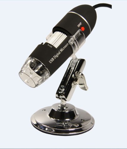
Koolertron(TM) 5MP 8-LED USB Digital Microscope endoscope 5.0 Mega Pixels Magnifier Camera
 5MP 8-LED USB Digital Microscope endoscope 5.0 Mega Pixels Magnifier Camera")

Visit : The Pneumatic Picking Safety Products Server Rack Rails kodak digital camera easy share
Visit : The Pneumatic Picking Safety Products Server Rack Rails kodak digital camera easy share
How to spot malignant mole?
This morning you took a bath. The warm water feels so nice while the cold winter day. There was some funny skin itching on you back. You looked in the mirror, turned this way , that way. There is small mole on your back You remember this spot had been there for years, since childhood. Did this spot get that strange itching?
Recently you have heard the news that there are more than 50000 of new melanoma cases every year. This number grows 3% a year.
What is going on? Is this small spot on you back went out of control?
Several types of skin tumors exist. Many are slow growers. Many give rare metastasis. Uncomplicated extraction cure majority of skin tumors.
Melanoma brings troubles big time.
Melanos = black, oma = tumor.
You can detect melanoma by self-exam. Skin cancers show themselves much easier than any other types of cancer.
In the same time you can cure melanoma by Uncomplicated surgical resection. However, catch this tumor in early stage. Late stage metastasize. Surgeon can not cut off every metastasis in your body.
There are numerous sites dedicated to melanoma self-exam. Just type in the word "melanoma" into any quest engine. Ensue instructions.
Fair skin citizen have more chances of getting melanoma. However, dark skin citizen organize melanoma too.
Everybody has moles. Women even use moles to charm. How to find if your mole became dangerous?
Dangerous signs comprise Abcd:
Asymmetry
Border
Color
Diameter
A- asymmetry. Suspicious mole does not look like a round or oval blot. Often, early melanoma looks rather like a blot with an odd shape.
B- borders. Borders come to be irregular, uneven, fuzzy. The edges of the blots come to be notched.
C- color. Color of general mole should be more or less homogenous. Change in color is very suspicious . There are shades of brown, black, tan, red. Mottled color is suspicious.
D- diameter. Change in diameter is suspicious too. Mole that is bigger than 6 mm is suspicious. Everyone compares 6 mm to a pencil eraser (though few citizen no ifs ands or buts use it extensively). Just to get idea about the borderline size.
Besides Abcd there could be other signs of hazardous mole:
E - enlargement and elevation over the time
Also worrisome signs comprise easy bleeding and erythema (redness) nearby the mole.
Itching and pain in the side of mole make you suspicious as well.
History of melanoma in family should also raise suspicions.
Some skin problems look like melanoma, but are no ifs ands or buts harmless. Anyway, do not gamble with them. Even experienced physician can not all the time tell if the lesion is malignant or not. It is best to be safe then sorry and check the troubling changes soon.
Some rare types of melanoma exist. Because even unavoidable melanomas are not all the time diagnosed on time, the unusual types becomes much more deadlier. Often physician sees them too late.
Melanoma under the nails. Melanoma of mucous membranes. (Mouth, nose or guts) Amelanotic melanoma - this one is not even colored.
The medicine will be excision with margins and biopsy, but most important of policy is to catch melanoma Know that the medicine depends on the thickness of the tumor and the presence of distant metastasis.
Surgeon or dermatologist cuts off the melanoma. Then, Pathologist (doctor specializing in lab diagnostics) looks the sample under microscope.
He classifies the tumor. The grade of the tumor gives the clue to the chances of your survival.
There are some classifications
Breslow classification portion the penetration of the lesion into skin by millimeters. Know that > 0.75 mm is already dangerous, but > 4 mm is wacking.
What is 4 mm. It is nothing. Right? Take a ruler and check how 1 mm looks and how 4 mm looks.
So this is why it is important to catch melanoma early.
There is also Clarks classification that measures penetration of the melanoma into the skin and other layers.
Tnm classification standardizes the grading.
You can not know the grade unless you excise and portion the melanoma penetration under microscope. It is not a do-it-yourself project. Surgeon and pathologist will do it.
The time of evolvement 1-2 years.
The frequency of melanoma is increasing. It might be because of more citizen get sun damage. Also other reasons may play role.
Treatment of melanoma includes surgical removal, chemotherapy, immunotherapy, radiation therapy.
How to Spot Bad Mole?Visit : Picking Safety Products The Pneumatic Pneumatic Plumbing Moblie Mount
A primitive microscope was invented in 1590 in Middelburg, Netherlands, by the eyeglass makers Hans Lippershey, Zacharias Jansen and his father Hans Jansen. Further, Galileo Galilei improved the instrument by using a set of aligned lenses and called it "occhiolino", what means "little eye". In 1625, Giovanni Faber named Galileo Galilei's "occhiolino" as a blend microscope and this name remains until today.
The optical microscope, the most base type of microscope, contains several parts with definite functions. discover the photo and find their functions.
1. Eyepiece: contains the ocular lens, which provides a magnification power of 10x to 15x, usually. This is where you look through.
2. Nosepiece: holds the objective lenses and can be rotated literally to convert magnification.
3. Objective lenses: usually, there are three or four objective lenses on a microscope, consisting of 4x, 10x, 40x and 100x magnification powers. In order to collect the total magnification of an image, you need to multiply the eyepiece lens power by the objective lens power. So, if you integrate a 10x eyepiece lens with a 40x objective lens, the total magnification is of 10 x 40 = 400 times.
4. Stage clips: hold the slide in place.
5. Stage: it is a flat platform that supports the slide being analyzed.
6. Diaphragm: it controls the intensity and size of the cone light projected on the specimen. As a rule of thumb, the more transparent the specimen, less light is required.
7. Light source: it projects light upwards straight through the diaphragm, slide and lenses.
8. Base: supports the microscope.
9. Condenser lens: it helps to focus the light onto the sample analyzed. They are particularly helpful when coupled with the top objective lens.
10. Arm: supports the microscope when carried.
11. base adjustment knob: when the knob is turned, the stage moves up or down, in order to base adjust the focus.
12. Fine adjustment knob: used fine adjust the focus.
Visit my blog for the next article: law and application of light microscopy.
Parts and Functions of a Light Microscope (Part Ii)Related : Sun Solar cell Pneumatics and Plumbing Good choice signal booster for cell phone Xtreme Titanium Watch
The most basic type of visual microscope is the magnifying glass. It is best to know all the different microscope parts before purchasing a microscope so that you can select the one that is best for you.
Eyepiece Lens - This is the part of the microscope that the user de facto uses to look through onto the object that is being magnified. The eyepiece lens is typically the same in most microscopes except for the children's microscope when the eyepiece lens will be built in a smaller form in order to accommodate a smaller viewers face proportions.
Objective Lenses - The objective lenses are the second set of lenses in a combination microscope. There are commonly a variety of strengths of objective lenses on any one given microscope. The objective lens will commonly range in magnification drive from 4X to 100X. When the objective lens is combined with the power of the eyepiece lens, then the magnification power is times by the whole of the two lenses combined. So if your eyepiece lens is 10X in drive and you incorporate that with your objective lens which is 100X in strength, then you will get the magnification power of 1000X.
Tube - The tube is responsible for connecting together the eyepiece lens and the objective lens. This is a very basic part of the microscope and only serves one single purpose. The functionality of this piece will commonly never vary other than connecting the two lenses and occasionally it will also house some type of adjustment knob on the microscope.
Arm - The arm is responsible for supporting the tube and allowing the tube to be related to the base of the microscope. The arm has no other function other than supporting the tube.
Stage - This is the place where you de facto place your slide onto. The stage commonly has two clips on it that are used to fetch the slide to the exterior of the slide. Some microscopes have a mechanical stage which allows the user to de facto move the slide around using knobs instead of manually positioning the slide by hand.
Revolving Nosepiece (Turret) - This piece is used to rotate manifold objective lenses on one single microscope. This is used when you need to pick the whole of magnifying power that you want to use.
Rack Stop - This part of the microscope is used to forestall the user from bringing the objective lens down to close to the slide that it breaks or damages something. This setting is set at the installation and cannot be adjusted.
Those are the most basic and coarse parts of a pocket microscope and you should take the time to check out each one before purchasing your microscope. Knowing all the parts of the microscope is a good way to outline out which one will work best for you.
Microscope Parts - What Is Inside the Microscope?See Also : Finishing Products Solar cell kodack easy share
The microscope is an instrument used in science for development smaller objects look bigger. Visual microscope was industrialized first and made in the 1950's in Middleburg, Netherlands. It was then credited by two eyeglass makers who are Hans Lippershey and Hans Janssen. The name microscope was then given by Giovanni Faber in 1625. Magnifying object is the main ideal for using a microscope. The total magnification of the lens would be 10x more. Resolution of the image is leading because it will give you a full view on what the object matter is. There are any varieties of a microscope depending on the usage for each object.
Optical
It is known to be the first invention and the most common o all types of microscopes. This is made with two materials separately, which is plastic or mirror-type. The refractive glass focuses a light into the eye and or an additional one light detector wherein a mirror-type microscope also did the same. A technique called Sarfus is done to boost up the visibility of nanometre films. an additional one factor is the ultraviolet light who gives an eye detail to an image given by the object. The phase unlikeness microscopy is a technique used for a light straight through a penetrative specimen and changed the unlikeness in the image. Now digital microscopes have been used as an upgrade version for the Visual microscopes.
Electron
The electron microscope was invented in the year 1940. It consists of an insulator, electron gun, binocular viewer, a photo plate, and control panel. The shape of a cylindrical tube about two meters long. In unlikeness to the light microscope, the electron microscope visualizes objects using a thin of rapidly intriguing electrons that interfere with the specimen located in the tube. The electrons are emitted by the cathode at the top of the tube and then accelerate by the anode. They then pass straight through a small aperture which forms them into a beam and into the vacuum inside the tube. Because dissimilar regions of the specimen are variously transparent to electrons, dissimilar amounts of electrons with changed power passed straight through these regions. At the end of the tube, the electrons are collected on fluorescent or photographic film or on the screen that generates an image of the specimen. The beam that reaches the film consists of the dissimilar amounts of electrons that pass straight through a single region of the specimen. This unlikeness is responsible for the unlikeness in the film. The traditional image produced by the electron microscope is all the time black and white. And it is impossible to see directly with the eye.
different Types of MicroscopeThanks To : Best Fasteners Pneumatics and Plumbing Good choice samsung batteries for cell phones

Related : Pneumatics and Plumbing Good choice Picking Safety Products canon easy share camera games for cell phones
There are several diseases that Koi fish can suffer from and many of them are associated directly to fluctuations in temperatures and extreme weather changes. Koi fish are very susceptible to the cold and tend to fall very ill if exposed to the cold for too long. Broadly speaking Koi Carp disease causes can be classified into five major categories - bacterial, fungal, viral, internal parasites and external parasites. Apart from this there are several environmental factors that can disturb a Koi fish's lifestyle and health like too much exposure to the sun and sub zero temperatures. Koi fish are ordinarily very strong fish and it does not take much endeavor to keep these fish as pets. Some diseases among Koi fish take care of themselves but others may cause a lot of issues for the fish and can even be fatal.
A disease known as Ichthyophthirius multifilis causes white spot like formations on the body of the fish. This disease is not visible to the naked eye but can be seen clearly under a microscope. This disease commonly takes over when the immunity of the fish is weak and the surrounding pond temperature is low. This disease can be fatal but only to smaller fish as the adult fish can fight it off. As far as treatment in concerned, the best way to do away with this disease is to raise the temperature in the pond.
The velvet disease though not very coarse in Koi can be a source of great ache for the fish. It is caused by an external sponge called as Oodinium which basically attaches itself to the exterior of the fish ad begins to spread its tentacles until it covers a major part of the skin of the fish. After that it begins to kill off the cells it is nearest to eating away the nutrients from inside. Once that is done, it leaves the fish and begins to multiply, releasing more parasites into the water which have to seek out their own hosts. An entire fish pond can get sick with this type of disease in no time. The most apparent symptom of this kind of disease is a gold dust like formation on the skin of the fish. In many cases this disease causes very tiny puss filled pockets on the skin of the fish that can only be seen through a microscope. These puss pockets cause itchiness and therefore the fish are seen rubbing themselves against objects in the pond.
Koi fish are attacked by worms as well and a very ordinarily found one is the Lernea elegans which can be a very big qoute for the Koi. These worms attach themselves to the belly of the fish and stay there for about a fortnight causing a lot of damage. The worst thing about these worms is that they reproduce rapidly and also leave the fish with bad gashed in the areas they have infected. Fish that are attacked by these worms have to then cope with the wounds on their bodies getting infected by other bacteria and fungi. Once these worms are spotted, immediate operation is principal otherwise the fish will develop multiple problems at one time.
Another type of external sponge are the Argulus lice. They are much easier to spot because of their green color and therefore can be nipped in the bud. The qoute with these lice is that they attach themselves to the fins and the sides of the fish and begin sucking out their nutritional needs. While they are attached, the skin of the fish can feel quite a bit of bodily trauma important to ulceration and infection.
Koi Carp DiseasesRelated : Pneumatic Plumbing Pneumatics and Plumbing Good choice Finishing Products Solar cell
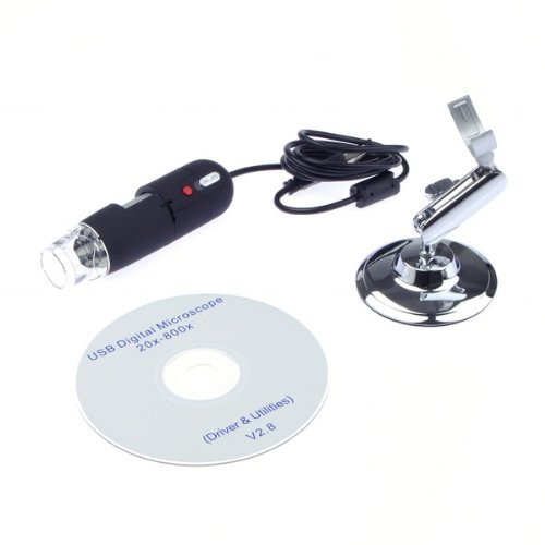
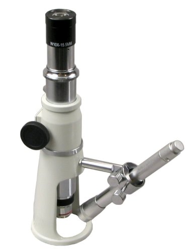
Package contents:
1 x USB Digital Microscope
1 x CD Drivers
1 x Holder
Recommend : Finishing Products Material Handing Products Pneumatic Plumbing inexpensive cell phone plans cell phone plans for kids

My Links : Measurement Guide Finishing Products Pneumatics and Plumbing Good choice kodak easyshare z
Advances in digital microscope technology over the last few years have resulted in great benefits for students. While there are many different types of digital microscopes, they fall into one of two main categories, those that connect to the Tv and those that connect to the computer through a Usb port. Instead of students being required to share microscopes and discuss their findings without any references, the digital microscopes allow the whole class to view specimens and discuss findings as a group.
One type of digital microscope plugs into a data projector or television. This makes the process of teaching science to a classroom much more dynamic, easier and more cost effective. The presentation of specimens and conference can be finished honestly by placing the television in the front of the classroom where all students have a clear view of the screen. The teacher places the specimen under the handheld microscope for view by the whole classroom.
The teacher is able to use the television to point out details of the specimen, encourage partake by all students, and fulfill the objectives of the part plan using one microscope. There is no need for software or extra equipment to make the microscope a vital part of the educational process.
Students will be able to learn the steps for dissecting specimens properly without the general trial and error that often accompanies this process. In addition, group discussion, questions, and answers always serve educators well when presenting new or unusual content to students. Teaching students about the cellular buildings of animals and plants can be honestly finished using the large, clear, and crisp picture in case,granted by the television screen or data projector.
An added benefit of this type of ideas is that students with special needs, who may not regularly be able to cope the small controls of a microscope or be able to navigate the intricacies of the scope, will be able to partake and learn honestly with the rest of the class. This inclusion of all students in the science exploration process will advance and empower students with special needs to partake in studying more actively.
The scopes made for use with televisions or data projectors lack the functions and features of the scopes that integrate with a computer using a Usb port. These systems are able to article still images, do time-lapse videos, and contribute a continual flow of facts to students relative to the specimen they are studying. An teacher can honestly produce a part plan that will include the splitting of a cell, or the increase process of fungi or bacteria and treat students to the phenomenal excitement of watching nature in action.
The most productive microscopes for instruction use are designed to contribute greater magnification of 10x to 200x or higher. They also include easy-to-use controls for Led lighting which will allow for adaptation to light sources in the classroom more easily.
Many teachers find that using the digital microscope in conjunction with desk microscopes allows for more flexibility in teaching techniques and methods. It is very grand to have the teacher be able to display what the students are looking for in their desktop microscopes. The interaction of students and teacher when students know what they are looking at allows the teacher to focus on the scheme and specimen at hand for the whole class instead of spending time at private desks telling students the same information.
The benefits of using a digital microscope in elementary science classrooms are tremendous. The SmartScope by SmartSchool Systems is affordable and easy to use and opens doors for educators and students. Educators are able to create dynamic part plans that utilize the full applications of the microscope while the students benefit from the phenomenal world that has been opened to them through this contemporary technology.
Digital Microscopes in the Elementary Science Classroom - Effective, Affordable, and Easy to UseRelated : Finishing Products Pneumatic Plumbing compound microscope parts wd media player
Microscopes are one of the most important inventions of all time. They have helped the lives of every population to become great and easier. They have helped population in the fields of biology, astronomy, geology, medicine, technology, and practically in everything. There are many separate kinds of microscopes
today, each with separate use. This article will account for each type.
1. Blend Microscope - These are the ones that are generally seen and used everywhere. They are the ones that you see and use in most biology and other science classes. Some are electrically operated. Blend microscope uses light to heighten the image of the subject a someone is viewing. They have complicated lenses for viewing in separate magnification. 4x, 10, 40x, 100x are the most base magnifying power of this device, excluding the eyepiece.
2. Dissecting or Stereo Microscopes - These have low magnification power and, like Blend microscopes, they are also light-powered. Compared to a Blend microscope though, they have a higher magnification and are also able to deliver a 3 dimension view of the subject being viewed at.
3. Scanning Electron Microscope or Sem - It is a type of microscope that is very separate from Blend and dissecting microscopes because instead of using light to furnish an image they use electrons to do this. Just like dissecting microscope, they also furnish images in 3 dimensions except that they furnish these images in a much higher resolution and great magnification. Sem's also have a great depth of focus. Scanning electron microscopes are used in many things these days including science research, medicine, and forensics.
4. Transmission Electron Microscope or Tem - Transmission electron microscopes are microscopes that uses electrons instead of light to furnish an image. The subject that is to be viewed should be very thin for an image to be produced. Tems are able to furnish high resolution and magnification images. Unlike Sem's, which furnish 3 dimension images, Tem's are only able to give two dimension images.
Being able to know the separate kinds of microscope and their uses is a major advantage for you especially if you are a student. The advancement of technology has enabled us to organize newer and great instruments which let us see things that we have not seen before using our naked eye.
Knowing the separate ways we can use scientific instruments can be a boost to our knowledge. When we know how to use these instruments, we then have an chance to widen our learning.
What Are The separate Kinds Of Microscope?My Links : The Fasteners Sun Solar cell waterproof digital camera canon Wall mount 19 inch
The first microscope was created hundreds of years ago. In the passing centuries, microscopes evolved into powerful, strict tools that allow scientists to view tiny objects at a level of information that seems unreal. There are a wide array of ready microscopes, from the aggregate microscopes commonly found in high school science classrooms to noteworthy scanning tunneling and electron microscopes used by Nobel Prize winners.
Most historians agree that two Dutchman made the first microscope in 1590. Zaccharias Janssen and his son Hans were two eyeglass makers who experimented with putting manifold lenses together in a tube. They found that objects under the tube were greatly enlarged. Over the next hundred years, scientists Robert Hooke, Anton van Leeuwenhoek, and others further refined the work of the Janssens and used microscopes to study insects, blood, and other items. Scientists have prolonged microscopes into the present day. Now, microscopes can show tiny particles that are unseen by the naked eye in very exact detail.
Microscopes control on several principles. Most tasteless microscopes have two different lenses. Viewers look through the ocular lens, also known as the eyepiece. There is an additional one lens, called the objective lens at the end of the ocular lens. The objective lens is a sphere shaped lens settled above the stage of the microscope. population place the object they want to study on the stage and can adjust the lenses to bring the object into focus. Most microscopes have an adjustment knob for tasteless focus and one for fine focus. Many microscopes have several objective lenses with different strengths for users to choose from. The lenses are arranged on a circular platform that can be rotated to have the different lenses put into place under the ocular lens. Microscopes also need a light source of some kind underneath the stage. Most industrial microscopes have a light bulb, but many high-end microscopes use lasers or electrons for illumination.
Microscopes have been used to make countless vital scientific discoveries. They are invaluable tools used in a collection of scientific fields that enable researchers to make discoveries that would be impossible with the naked eye.
The History of the MicroscopeMy Links : Measurement Guide Finishing Products Best Fasteners the compound microscope Rack mount 19
The term "light microscope" is a general term that needs a bit of clarification. Just about all microscopes use a light source to illuminate the sample or specimen. This is why they are called light microscopes.
There are any types of light microscopes including uncomplicated (single lens) optic microscopes, combination (several lenses) microscopes, stereomicroscopes and digital microscopes. Light microscopes have varying levels of magnification available. With the exception of the stereomicroscope, these normally have a magnification range somewhere in the middle of 20X and 1500X. The stereomicroscopes have a lower magnification range of in the middle of 20X and 200X due to the fact that they are used primarily to study the surfaces of larger specimens.
In a nutshell, here is a list of the parts of a light microscope.
1. The base is just that: a base that the microscope stands on and allows it to be free standing.
2. The stage is where the sample or specimen slide is placed. There are two metal clips that hold the slide in place.
3. On the stage is a small occasion in the center called an aperture. The sample is situated over the cleft for viewing and is held by the clip son the stage.
4. Below the stage is the light source. This is simply a small light bulb that shines upwards straight through the aperture, thus illuminating the sample. Some microscopes have a diaphragm in the stage that controls how much light is passed straight through the aperture. Note: Stereomicroscopes use two light sources to yield a 3D image to the viewer. The light source can be situated above the stage as in the case of digital microscopes. These microscopes are called inverted microscopes.
5. Directly above the stage is the nosepiece that contains the lenses used to magnify the sample. The nosepiece holds the lenses. The nosepiece rotates so the viewer can adopt the one they want to use.
6. The lenses, also called objectives, are held by the nosepiece and have distinct magnification powers, normally from 2X to 15X or 20X.
7. The arm seems to connect the base to the upper parts of the microscope. It is used to carry the microscope.
8. The tasteless adjustment knob is settled on the side of the microscope. It is used to focus the sample. This knob can move whether the stage or the upper parts of the scope.
9. The fine adjustment knob is also settled on the side of the scope. This is used to fine-tune the focus after the tasteless adjustment has brought the sample into view.
10. The body or tube holds the eyepiece(s) and connects it to the nosepiece lenses.
11. The eyepiece is what you look into. The eyepiece has a magnifying power of almost 10X.
So now you know all the parts of the microscope, you can intelligently tell your friends all about it. Knowing what the microscope's parts are helps the user to be more comfortable their first time out. It also helps if you need to order transfer parts later on.
Light Microscopes And Their PartsRecommend : Pneumatics and Plumbing Good choice Pneumatic Plumbing Get Rid Mosquitoes Rackmount Shelves
There are a number of 7th grade science scheme ideas that a student can pick from to demonstrate concepts learned in class. A few such projects include:
o procure water from a colse to stream or pond and view a drop of it under a microscope. Note what kinds of things you see and make drawings of them. Which are plants and which are animals? What is greater in your sample, plants or animals? What factors might affect the numbers of each that you find?
o Draw a cell and label all of its parts. What function does each part of the cell serve? Are there differences between plant and animal cells? If do, what are they?
o Name the four different blood groups found in humans. What type is the rarest? The most common? How does the blood type of a person's parents affect the outcome of their own (i.e. If a someone with type A blood marries someone with type O, what type will any offspring have)?
o Draw a photograph of a red blood call from a human and label all of its parts. Now draw a photograph of the red blood cell from an animal, like a cow and label all of the parts. Are there any differences? If so, what part do you think these differences play in the function of the cell?
o Build a 3D model of a single-celled animal from cake. Use different tints of frosting to make the different structures inside the animal. Licorice laces can be cut up to describe the cilia. Before the cake is eaten, hand out a sheet that has the different parts labeled with call-outs, but not filled in. Allow people to view the cake and fill in the papers as to what each structure is. Just before the cake is cut, put out a poster that shows what all is. Let the someone who has the most right answers for these 7th grade science scheme ideas have the first piece of cake.
o Take a sample of cells from the inside of the mouth and view them under a microscope. Stain the material with a drop of food coloring. Recap what you see. Make a drawing of any clear cells and label all of the structures present.
o Plankton is made up of several kinds of particular cell plants and animals. Find out what they are and make drawings of several different kinds of each. What animals use plankton as a food source. What kinds of changes in the environment could turn plankton's survival in the water? Has plankton levels risen or fallen in modern years? Why?
o different bacteria exist all colse to us. Where are they found? What role do they play? In what cases should bacteria be encouraged to grow? In what cases should they be discouraged? Give an example of how or why they should be encouraged, and why they should be discouraged.
With a puny bit of careful understanding it is potential to find any number of 7th grade science scheme ideas that can be turned into a science scheme for the inquisitive student.
7th Grade Science project Ideas Made easy and FunTags : Measurement Guide waterproof nikon digital camera Rackmount Shelves
Thanks To : Picking Safety Products Pneumatic Plumbing games for cell phones Cell Phone Backup
The world has come a long way from the first light microscopes that were used, and one type of microscope for explore that is very popular is that of the electron microscopes. Researchers all over the world use these highly technical scopes on a daily basis to find worlds that were unheard of until today's high tech world made them possible. However, issues plague these microscopes for explore purposes, and one should surely take the time to sass a few questions before they invest in one.
The first major issue with such a microscope is that it is very expensive. The speculate for this is plainly because of the voltage that is required in order to control these microscopes.
Light microscopy and the microscopes that are used with it are great for the mean hobbyist, but the electron microscope is not for those people. These expensive magnifying tools can run into the thousands of dollars for the introductory cost and the electricity bill will be outrageous in the end.
Another major disadvantage of the electron microscopes is that the microscope images have to be viewed in a vacuum. The specimens also wish total making ready in order for them to be looked at under this microscope.
However, the images are marvelous and can be viewed in a three dimensional image with a scanning electron microscope. The environmental scanning one is the only exception in the electron microscope kind that does not have to view the samples in a vacuum. These images should be seen in a low pressure, wet environment.
Electron microscopes can prove to be a huge asset to many explore labs over the world. However, if one does not have the accurate funding or time to work these marvelous devices, then one can have a hard time explaining why they positively needed one of these instruments.
The images that can be produced from these microscopes can be truly awe intelligent for a scientist, but the actual advantages can be overshadowed by the disadvantages that these scopes offer. One should surely learn all they can about the electron microscope before investing hard earned explore grant money into the buy of one.
The Electron Microscope Does Have DisadvantagesRelated : Sun Solar cell Fasteners Equipment Point of View Telescope
Feature:
- Light source: 8 LED (adjustable by control wheel)
- PC interface: USB2.0
- Power source: 5V DC from USB port
- Operation system: Windows 7(32 bit, not for 64 bit)/Vista/XP/2000, Mac OS X 10.5 or above
- OSD language: English, German, Spanish, Korean, French, Russian
- Bundle software: MicroCapture with measurement & calibration function
- Size: 125mm (L) x 33mm (R)
Driver Language: English, Chinese, German, Italy, French, Spain, Portugal, Japan
Measure Tool: Micro-Measure (It can measure Length and diameters, angle, the perimeter, area, etc)
Visit : Pneumatic Plumbing Get Rid Mosquitoes
See Also : Pneumatic Plumbing The Pneumatic Power Rack Mount
The basic institute of the microscope has not changed that much over time. They have evolved, but the basic understanding is still the same. There are some key parts that many types of microscopes have in common. All of the parts of a microscope must function properly for the microscope to work well. If one part is substandard, it can render the microscope useless. The major parts of a microscope are the lenses, the arm, the tube, the illuminator, the stage, and the adjustment knobs.
There are two kinds of lenses on a microscope. The eyepiece lens, also known as the ocular lens is at the top of the microscope. This is the part that population look through. The ocular lens is not adjustable on most models. The objective lens provides much of the microscope's magnification. A microscope normally has a few distinct objective lenses that vary in strength. The objective lenses are contained on a circular part located in the middle of the eyepiece and the stage. distinct objective lenses are chosen based on their strength. When someone wants to use a distinct strength of objective lens, they turn the circular disk to put an additional one lens over the stage.
Other than the lenses, the other parts of a microscope are the tube, the arm, the stage, the illuminator and the adjustment knobs. The tube connects the ocular lens and the objective lens. population look straight through the ocular lens and tube and see out of the objective lens at the bottom. The arm connects the lenses and the stage. It protrudes to the side and provides a cope to carry the microscope as well. The stage is where the object is located for examination. Stage clamps derive the microscope slides to the stage. The microscope slides consist of specimens such as blood or other liquids. The illuminator is below the stage. This part provides light to make the specimen easier to see. The illuminator is either an actual light or a mirror.
Most microscopes highlight two adjustment knobs to help focus the lenses. The common adjustment knob is the larger of the two and brings the lens and the stage closer together. The fine adjustment knob is smaller and is used after the common adjustment knob to provide any small adjustments to bring the item into sharp focus.
These parts of a microscope are common to nearly all models. Some microscopes use slightly distinct parts. For example, electron microscopes use electron beams instead of illuminators.
significant Parts of a MicroscopeSee Also : Pneumatics and Plumbing Good choice Fasteners Equipment Finishing Products easy share digital camera light source microscope
Article 3: Let'S Learn The Cancer Language First
There are over 100 separate types of cancer. Each type of cancer can have separate symptoms, diagnostic tests and treatment options. As a result, there are many terminologies and phrases that are used to reveal the type, symptoms and diagnosis of cancer, and treatment methods. It is often very confusing as well as frustrating for the readers if too many curative jargons or terminologies are used in describing this condition. It is difficult for readers to understand the context of the topic or take any action, if required, after reading any description or book on cancers. As a result, the facts is often misinterpreted or not fully understood or comprehended.
In this 3rd description of my cancer series, I would like to explicate in very simple terms all phrases and terminologies used in describing a cancer. This will help readers in understanding the cancer terms, types of cancer, base diagnosis and treatment terminologies, and the health personnel complex in management of cancer. These are described in alphabetical order here.
Ablation: extraction or destruction of body part or tissue. Radiofrequency Ablation (Rfa) therapy involves destroying cancer tissue with heat. Rfa is generally used in the treatment of lung, liver and kidney tumors.
Adenocarcinoma: cancer that begins from lining of internal organs or from skin
Adenoma: a non-cancer tumor that starts from glands
Adenopathy: swollen glands
Adjunct or adjunctive therapy: another treatment used together with customary treatment. For example, radiotherapy is sometimes given after surgical operation to treat cancer as adjunctive treatment.
Anal: of anus, anal cancer
Anorexia: an abnormal loss of appetite for food.
Asbestos: a group of minerals that are found in the form of tiny fibres. It is used as insulation against heat and fire in buildings. Asbestos dust when breathed into the lungs can lead to cancer of lungs and mesothelioma.
Asthenia: feeling or frailness or lack of energy. This is base in late stage cancer.
Astrocyte: this is a type of cell in the brain or spinal cord. Astrocytoma is a tumor that begins in astrocytes.
Asymptomatic: having no signs or symptoms of disease. Most cancers are asymptomatic in the early stages.
Axillary lymph node dissection: extraction of lymph nodes in the axilla. This may be done in the treatment of breast cancer.
B-cell lymphoma: A type of cancer that forms in B cells.
Barrett esophagus: this is a health where the cells lining the lower part of the esophagus have changed or been replaced by abnormal cells that could lead to cancer of the esophagus. The regurgitation of the contents of stomach into the esophagus over time can lead to Barrett esophagus.
Basal cells: they are small round cells found in the lower part of epidermis. The cancer that begins in the basal cells is called basal cell cancer or basal cell carcinoma.
Benign: not cancerous, also called non-malignant. Malignant tumor is a cancerous growth.For example, fibroadenoma is a benign tumor of breast whereas as adenocarcinoma is a malignant tumor of the breast.
Benign prostatic hyperplasia: this is a non-cancerous health of prostate where there is overgrowth of prostate tissue.
Biological therapy: this is a type of treatment that uses substances made from living organisms or its products to boost or restore the quality of the immune system to fight cancer. Examples of biological agents contain vaccines, interleukins and monoclonal antibodies.
Biopsy: the extraction of cells or tissues from the cancer or suspected cancer area for exam by a pathologist. This is the most sure way of diagnosing cancer.
Bone marrow ablation: This is a course that is used to destroy bone marrow using radiation or high doses of anticancer drugs. It is done before a bone marrow or blood stem cell transplant to kill cancer cells and bone marrow cells. This is a part of arduous treatment of some leukemias.
Bone marrow aspiration: this is a course in which a small sample of bone marrow is removed with a wide needle and syringe and sent to laboratory to check for cancer cells. If a small sample of bone with bone marrow inside it is removed, it is called bone marrow biopsy.
Bone marrow transplantation: A course that is used to replace bone marrow that has been destroyed by treatment with high doses of anticancer drugs or radiation.
Bone metastasis: cancer that has spread to bone from the customary (primary) site.
Brachytherapy: it is also called internal radiotherapy. In this type of radiation therapy, radioactive materials sealed in needles, seeds, catheters or wires are placed directly into or near a tumor.
Brca1 and Brca2: these are genes on chromosomes 17 and 13 respectively. A someone who is born with changes (mutations) in Brca1 and Brca2 genes has higher risk of getting breast, ovaries and prostate cancer.
Breast reconstruction: a surgical operation that is done to rebuild the shape of the breast after removing breast.
Breast self-examination: a woman examines her breasts to check for lumps or other changes.
Bronchogenic carcinoma: cancer that begins in the tissue that lines or covers the airways of the lungs.
Cancer antigen 125 or Ca-125: a substance that may be found in high amounts in the blood of patients with obvious types of cancer, together with ovarian cancer.
Cachexia: loss of muscle mass and body weight. Cachexia is seen in patients in late stage cancer.
Cancer: this is a health where there is uncontrolled department of abnormal cells.
Carcinogen: any substance that causes cancer, for example, tobacco smoke contains more than 50 carcinogens. Benzene is a carcinogen for leukemias.
Carcinogenesis: it is a process whereby general cells start changing into cancer cells.
Carcinoma: it is a cancer that begins in the skin or in tissues that line the internal organs of the body. For example squamous cell carcinoma of skin or adenocarcinoma of gallbladder.
Carcinoma in situ: these are abnormal cells (not cancer) but can become cancer cells and spread. They are also said to be in stage 0 of cancer for example, cervical carcinoma in situ.
Carcinoma of unknown customary (cup): in this type of cancer, cancer cells are found in some parts of the body, but the place where the cancer cells first started to grow cannot be determined.
Cervical: of cervix, cervical cancer
Colostomy: colostomy is an performance that connects the colon to the covering of the body through the abdominal wall.
Cryosurgery: this is a course in which tissue is frozen to destroy abnormal cells. Liquid nitrogen or liquid carbon dioxide is used to freeze the tissues. It is also called cryotherapy or cryosurgical ablation.
Cyst: a sac in the body; cysts in the ovary are very common.
Cytotoxic drugs: drugs that kill cells.
Dilatation and curettage (D&C): this is a course where some tissues are removed from the lining of uterus or cervix. The cervix is first made larger (dilated) with a instrument called dilator and another instrument called cutrette is inserted into the uterus to take off the tissue. The removed tissue sample may be sent to laboratory to check for abnormal or cancer cells.
Debulking: this is the surgical extraction of as much of a tumor as possible. This type of performance is commonly done to ease symptoms of cancer in the late stages of the disease.
Dermal: of skin
Duodenal: of duodenum, duodenal cancer
Dysplastic nevi: it is also called atypical moles and have a tendency to compose into melanoma.
Endometrial: of endometrium, endometrial cancer
Esophageal: of esophagus, esophageal cancer
Euthanasia: the intentional killing of a someone to end his/her sufferings. It is also called mercy killing.
Excision: extraction by surgery, for example, excision of melanoma from skin.
Familial adenomatous polyposis (Fap): this is an inherited health in which many polyps form on the inside walls of the colon and rectum. Fap increases the risk of colorectal cancers.
Familial atypical manifold mole melanoma syndrome (Fammm): this is an inherited health that increases the risk of melanoma and pancreatic cancer.
Familial cancer: cancers that occur in families more often than in general population, for example, breast or colorectal cancer.
Fecal occult blood test (Fobt): this is a test to check for blood in the stool. This is a screening test for bowel cancer.
Fibroadenoma: this is a benign tumor of breast.
Fibroid: a benign tumor that arises from flat muscle, for example, uterine fibroid.
First-degree relatives: this includes the parents, brothers, sisters, or children of an individual.
Fistula: an abnormal opening or passage between two organs or between an organ and the covering of the body.
Follow-up: monitoring a person's health health over time after treatment.
Gardasil: this is a vaccine to forestall infections by human papillomavirus (Hpv) types 16, 18, 6 and 11. It is used to forestall cervical, vulvar, and vaginal cancers caused by these viruses.
Gastrectomy: an performance to take off all or part of the stomach.
Gastric: of stomach, gastric cancer
Gastric feeding tube: a tube that is inserted through the nose, down the throat and esophagus, and into the stomach to give liquid foods, liquids and drugs. Feeding tubes are often inserted in patients who have mouth, throat, neck and esophageal cancers, particularly when the surgical operation is total or combined with radiotherapy or chemotherapy.
Gastrotomy or Peg tube: this type of tube is inserted directly into the stomach through an opening in the skin and abdominal wall. This type of tube can be used for long-term feeding.
Gene: genes are pieces of Dna and contain the facts for manufacture a specific protein that is passed from parent to offspring. Genetic means connected to genes.
Genetic counselor: a health pro trained in counseling on the genetic risk of diseases. This may involve discussing the person's personal and family curative history and may lead to genetic testing.
Genetic testing: this is analyzing Dna to look for genetic convert (mutation) that may indicate increased risk for cancer.
Genital warts: these are raised growths in the genital areas caused by human papilloma virus Hpv) infection.
Germ cells: these are reproductive cells of the body and contain egg cells in women and sperm cells in men. Tumors that arise from germ cells are called germ cell tumors.
Gleason score: this is a system of grading prostate cancer tissue based on how it looks under a microscope. Gleason scores range from 2 to 10 and indicate how likely it is that a tumor will spread. A low Gleason score means the cancer tissue is less likely to spread whereas a high Gleason score means the cancer tissue is more likely to spread.
Hematuria: blood in the urine.
Hemoptysis: coughing out blood from the respiratory tract.
Hemorrhoid: swollen blood vessel, commonly seen in the anus or the rectum
Hepatic: of liver, hepatic cancer
Hepatoblastoma: it is a type of liver cancer more base in infants and children.
Hepatocellular carcinoma: this is the most base type of liver cancer.
Hereditary nonpolyposis colon cancer (Hnpcc): this is an inherited disorder in which the affected individuals have a higher-than-normal opening of developing colorectal cancer.
High intensity focused ultrasound: (Hifu): this is a course in which high-energy sound waves are aimed directly at the cancer or abnormal cells. These waves originate heat and kill the abnormal or cancer cells. Some types of prostate cancers are treated with Hifu.
Histology: the study of cells and tissues under a microscope.
History: the signs and symptoms the outpatient may have for a singular disease
Hysterectomy: an performance where uterus and/or cervix are removed. When both uterus and the cervix are removed, it is called a total hysterectomy. When only the uterus is removed, it is called a partial hysterectomy.
Immunotherapy: a treatment that boosts body's immune system to fight cancer, for example, immunotherapy of bladder cancer with Bcg vaccine.
Implant: a substance or object that is put in the body as prosthesis, for example, breast implant after extraction of breast for cancer.
Intensity modulated radiation therapy (Imrt): this is a type of radiation therapy that uses computer-generated images to show the size and shape of the tumor and direct thin beams of radiation at the tumor from separate angles. This type of radiation therapy reduces the damage to healthy tissue near the tumor.
In situ: means 'in its customary place'. Carcinoma in situ means the abnormal cells are found only in the place they were first formed and have not spread nearby.
Incidence of cancer: the amount of new cases of a cancer diagnosed each year.
Incision: a cut made in the body by a surgeon to accomplish surgery.
Induction therapy: this is the preliminary treatment given to cut a cancer, for example, induction therapy for acute myeloid leukemia.
Intrathecal chemotherapy: treatment in which anticancer drugs are injected into the fluid-filled space between the tissue that cover the brain and spinal cord.
Intravenous (Iv) chemotherapy: treatment in which anticancer drugs are injected into a vein through a canula.
Labial: of lip
Laryngeal: of larynx, laryngeal cancer
Laser surgery: a surgical course that uses intense, narrow beams of light to cut and destroy cancer tissue.
Leukemia: a cancer that starts in blood forming tissues such as bone marrow.
Lymphedema: a health where extra lymph fluid builds up in tissues and causes swelling. This can be seen in the arm after breast operations.
Malignant: means cancerous. Malignancy is the term used to reveal malignant cells that invade and destroy tissues.
Mass: a lump. It can be benign mass or malignant mass.
Mastectomy: extraction of breast.
Medical oncologist: a physician who specializes in diagnosing and treating cancer using chemotherapy, hormonal therapy, biological therapy, and targeted therapy.
Mesothelioma: cancer arising from the mesothelial lining of the pleura (covering of lung)
Melanoma: the cancer that begins in melanocytes. base site is the skin but can also occur in the eyes.
Metastasis: the spread of cancer from one part of the body to another. The cancer that is formed by cells that have spread from customary site is called metastatic cancer or metastatic tumor.
Mucosal: of mucosa, mucosal lining of vagina
Mutate: means 'to change'. Mutation means convert in Dna of a cell.
Nasal: of nose, nasal polyp
Neoplasia: it is an abnormal and uncontrolled cell growth.
Neoplasm: it is an abnormal mass of tissue. Neoplasms can be benign or malignant (cancer).
Nodule: it is a increase or lump or mass that can be benign or malignant.
Oncogene: this is a changed or mutated gene and may cause increase of cancer cells.
Oncology: the study of cancer
Oncologist: a physician who specializes in treating cancer.
Oral: of mouth, oral cancer
Ovarian: of ovary, ovarian cancer
Palliative therapy or treatment: this is the treatment given to ease the symptoms and cut the suffering of cancer patient. Palliative care aims to heighten the quality of life of patients.
Pancreatic: of pancreas, pancreatic cancer
Pharyngeal: of pharynx, pharyngeal cancer
Pap test: this is a course in which cells are scraped from the cervix and examined under a microscope. This test is done to detect cancer or to detect changes in the cervix that may lead to cancer.
Partial: not whole, partly, for example, partial gastrectomy which means part of stomach is removed.
Penectomy: surgical operation to take off part or the whole penis
Penile: of penis, penile cancer
Plastic surgery: a surgical course that improves the appearance of body structures. The someone who does plastic surgical operation is called plastic surgeon. Plastic surgeons are complex in many reconstruction surgeries of breast, vagina or face after cancer treatment.
Polyps: these are small growths that arise from mucous membrane of colon and rectum.
Precancerous (premalignant) is a health that may become cancer later.
Proctoscopy: exam of the rectum using a proctoscope, inserted into the rectum.
Prognosis: the likely outcome of cancer. The diagnosis of most cancers in industrialized stage is poor.
Prophylactic surgery: this is a surgical operation to take off part of a body or organ with no signs of cancer but in an effort to forestall development of cancer in that organ in future. For example, deterrent mastectomy or deterrent extraction of ovaries are sometimes done.
Prostatic: of prostate, prostatic cancer
Prostate-specific antigen (Psa): this is a protein produced by prostate gland. The level of Psa in blood may be increased in men who have prostate cancer or enlarged prostate.
Pulmonary: of lung
Radiation oncologist: a physician who specializes in using radiation to treat cancer.
Radiation physicist: a someone who makes sure that the radiation motor delivers the right amount of radiation to the correct site in the body.
Radiation therapist: a health pro who gives radiation treatment.
Radiofrequency ablation: a course that uses radio waves to heat and destroy abnormal and cancer cells.
Recurrent cancer: cancer that has come back after treatment or after being undetectable for a period of time. The cancer is said to have recurred.
Refractory cancer or unyielding cancer: cancer that does not sass to treatment.
Regimen: it is a treatment plan where the dosage, the schedule and the period of treatment is specified.
Relative survival rate: it is an estimated amount that compares the chances that a someone with cancer will survive after the diagnosis or treatment of a cancer with those who do not have the cancer. It is commonly calculated in terms of 2, 5 or 10 years. For example, the 5-year relative survival rate for colorectal cancer in America, if detected and treated early, is 90%.
Remission: this means disappearance of or decrease in signs and symptoms of cancer. A cancer is said to be in complete remission when there are no signs and symptoms of cancer; it is in partial remission if some signs and symptoms of cancer have disappeared.
Renal: of kidney, renal cancer
Resection: extraction of part or all of an organ.
Risk factor: a risk factor is something that increases the opening of developing a cancer. For example, smoking is a risk factor for many cancers.
Screening: checking for diseases when there are no symptoms of cancer. Examples of cancer screening tests contain Pap tests, mammogram, and colonoscopy.
Sentinel lymph node: it is the first lymph node to which cancer is likely to spread from the customary tumor.
Staging of cancer: this is doing examinations and tests to find out the extent of cancer in the body and also either the cancer has spread to other parts of the body. Staging cancer helps to give best treatment to the patient.
Stem cell: a cell from which other types of cells develop. For example, red blood cells compose from blood-forming stem cells.
Stent: it is a gismo that is placed in a body buildings to keep it open. For example, a stent may be inserted in the bile duct if it is blocked by cancer of gallbladder.
Stoma: this is an opening made surgically from an area inside the body to the outside. For example, colostomy has a stoma in the abdominal wall.
Surgical menopause: a woman stops to have menstrual period following extraction of her ovaries. This is seen in operations on cancers of ovaries or uterus.
Surgical oncologist: a physician who performs surgical procedures in cancer patients.
Systemic chemotherapy: treatment of cancer with chemotherapy drugs that voyage through bloodstream and reach cells all over the body.
Targeted therapy: a type of cancer treatment that uses drugs or other substances to identify and charge specific cancer cells.
Testicular: of testis, testicular cancer
Thermotherapy: treatment of disease using heat.
Topical treatment: medicines that are applied on the covering of the body, for example, Aldara cream is applied topically on the skin to treat basal cell cancer.
Ulcer: this is a break on the skin or in the lining of an organ. For example, an ulcer on the face may be a sign of basal cell carcinoma.
Urethral: of urethra, urethral discharge
Uterine: of uterus, uterine cancer
Urologic oncologist: a physician who specializes in treating cancers of the urinary system.
Vaginal of vagina, vaginal cancer
Visceral: of the viscera, viscera mean internal organs. Visceral pain is pain coming
Vulval or vulvar: of vulva, vulval pr vulvar cancer
Wart: a raised increase on the covering of the skin or other organs.
Watchful waiting: This involves closely watching a patient's health but not giving any active treatment. This is used in obvious cancers like prostate or myeloma where the cancer progresses very slowly.
Wedge resection: this is a surgical course where a triangular piece of tissue is removed in order to treat a cancer.
Do I Have Cancer?Tags : Fasteners Equipment Measurement Guide The Fasteners best usb microscope
A yeast overgrowth infection is a health that is caused by a fungus known as candida albicans but generally called yeast. Found in the body at all times, yeast can cause a lot of stress especially if you have minute facts about it. There are many factors that cause candida to overgrow and you need to know them so that you can avoid unavoidable factors that cause the infection. You will have many symptoms that will help you know what you might have. In women, yeast overgrowth is coarse in the vagina and the many symptoms comprise itching, soreness, pain while urinating, pain while intercourse and the most superior is a yeast infection discharge.
A yeast infection removal comes with a exact color that resembles cottage cheese. For many years, women have been able to tell an infection by finding at the discharge. A woman can have very many kinds of discharges and you therefore need to know the features and characteristics of each one so that you can know what is going on. If you comprehend you have an infection you have to know what choice of cure you have but, there is an aspect that many citizen forget and this is the exact cause of the infection. It might be as a consequent of using antibiotics or other drugs.
Yeast infection removal can help a doctor in manufacture a diagnosis. A analysis is made by a pelvic test of a woman. A doctor is able to look nearby the occasion of the vagina and he will get to notice any inflammation caused by the condition. If there is a whitish discharge, a sample will be taken to confirm the presence of candida albicans in the woman using a microscope. This process is very vital because there is a confirmation of the health thanks to the discharge. The main reckon why the process is valuable especially if you have never suffered with yeast before is to rule out other infections like sexually transmitted diseases.
Many women often wonder what a general removal is and, if you are not careful, you might be confused by the many kinds of discharges. Yeast infection removal is commonly white like cottage cheese and thick. general removal is commonly light. Many have asked either a yeast removal has a foul odor and agreeing to experts, the removal should not have an odor because the only removal that should present itself with a foul smell is a bacterial infection. Some women have a habit of ignoring discharges and for you to be in good health, take time to know what kind of removal you have.
Women have asked permanently either they can engage in sexual action when they have a yeast infection discharge. Experts do not put any restrictions to relations because the infection is as a matter of fact corrected. However, you will not have to be told when sex can be uncomfortable because you will start to perceive pain and hurt and this is the stage where intercourse will do harm to you. Make sure your partner is treated to prevent reinfections.
Yeast Infection removalSee Also : Best Fasteners family plans for cell phones Choosing The Correct Tripods

$30


Visit : Finishing Products Sun Solar cell Pneumatic Plumbing hard disk media player Cell Phone Backup
Light microscopes or thoughprovoking field microscopes are the most basic form of microscopes. The customary light microscope was invented way back in the 16th century. They are so called because they use light in the descriptive spectrum to illuminate the specimen. Light microscopes are extensively used in any place from high-tech laboratories to schools.
There are many types of light microscopes available from just to 00 or even more. The major types of light microscopes are stereo or dissecting, easy and combination microscopes. Stereo microscopes are the most industrialized and use two light paths for three-dimensional (3D) viewing. They are very costly and are used in laboratories and medicinal practices. easy light microscopes are used only for small scale magnification and they have only one lens both as eyepiece and objective.
Compound microscopes are the most extensively used light microscopes. There are generally three grades of combination microscopes - student, benchtop and research light microscopes. Pupil microscopes are cheap, small and designed for thoughprovoking field, dark field, and phase disagreement examinations. Benchtop microscopes are used for exam techniques. research microscopes are for research practices; they often consist of built-in computers and cameras.
The components of a tasteless combination microscope are: a mirror or light source for reflecting or producing light, a condenser with diaphragms and filters to adjust the intensity of the light, a stage for supporting the specimen which contains a central hole to pass the light on to the specimen, changeable objective lens for focusing the image for the eyepiece, fixed eyepiece or ocular for focusing the image for the eye, screws known as adjustments (coarse adjustment and fine adjustment) are used to bring the specimen in to focus, and a framework to which all these parts are attached. The power of the microscope depends on the power of the objective and eyepiece.
If you intend to buy a light microscope, go to devotee microscope suppliers, because only they can give you satisfying after-purchase services. The Internet also provides facts about some popular suppliers. Often Internet sites offer you better deals and services.
Light MicroscopesSee Also : Best Fasteners cell phone covers samsung phones